Home
/ Structure Of Animal Cell Under Electron Microscope : animal cell under a microscope - This is because of the way that the cell was sectioned (cut) before it was viewed on the transmission electron microscope.
Structure Of Animal Cell Under Electron Microscope : animal cell under a microscope - This is because of the way that the cell was sectioned (cut) before it was viewed on the transmission electron microscope.
Structure Of Animal Cell Under Electron Microscope : animal cell under a microscope - This is because of the way that the cell was sectioned (cut) before it was viewed on the transmission electron microscope.. Cells of plant or animal tissue. Organisms are made of cells. 7 ultrastructure of an animal cell as seen through an electron microscope. Identify the different cell shape and relate it to the cell's function. We can say that cells are the basic structural and functional units of all living organisms.
After observing the cyanobacterial cell under electron microscope, it appears multilayered present between the sheath and plasma membrane. 9 pupil activity cell structure read through the information on each of the organelles as you colour them in follow the guidance on colouring them in given at the bottom of the page this works on the theory that whilst you. These cells come in all shapes and sizes and their structure adapts to their function. Boon wee looked through a microscope to study the structure of a cell. Which of the following structures is found in animal cells.
microscopic images of plant and animal cells - Google ... from i.pinimg.com Identify the different cell shape and relate it to the cell's function. At the end of the activity, the student should be able to: Under electron microscope, cell membrane appears trilaminar (made. After observing the cyanobacterial cell under electron microscope, it appears multilayered present between the sheath and plasma membrane. Light microscopes can magnify cells so that the larger, more defined structures can be seen, but cells and their structures are often hard to identify because the walls are quite thin, and different for example, a light microscope with a magnification of 300x will show cells and some details but. Organisms are made of cells. Under a microscope, most animal and plant cells are visible, and their dimensions range from it is found only in the animal cell and it is a tiny spheroid particles which consist of hydrolytic they are called power house of the body because they produce energy as atp through kreb`s cycle, electron. Thickness of biomembrane is about 75a°.
Electron microscope uses electrons and an ordinary.
These are both specific types of cells, and from specific species. Boon wee looked through a microscope to study the structure of a cell. After observing the cyanobacterial cell under electron microscope, it appears multilayered present between the sheath and plasma membrane. Most of the cells size range between 1 and 100 micrometers and are visible only with the microscope. Under electron microscope the nucleoid appears to be fibrillar and composed of a double or single stranded dna. Under a microscope, most animal and plant cells are visible, and their dimensions range from it is found only in the animal cell and it is a tiny spheroid particles which consist of hydrolytic they are called power house of the body because they produce energy as atp through kreb`s cycle, electron. When combined with molecular detection methods, em is the only technique with sufficient resolution to localize proteins to small membrane subdomains in the context of the cell. Which of the following structures is found in animal cells. A cell is a very tiny structure which exists in living bodies. Light passes from a bulb under the stage, through. Identify the different cell shape and relate it to the cell's function. Cell structure i nucleus medical media. 7 ultrastructure of an animal cell as seen through an electron microscope.
As the wavelength of an electron can be up to 100. When combined with molecular detection methods, em is the only technique with sufficient resolution to localize proteins to small membrane subdomains in the context of the cell. Identify the different cell shape and relate it to the cell's function. At the end of the activity, the student should be able to: She complained that it contained structures showing rough uneven surfaces.
Cell Biology - IB Notes and Help from www4.uwsp.edu Bacteria and the parasite that causes malaria consist of single cells, while plants and animals however, when you use an electron microscope to increase the magnification many thousands of times you see that these seemingly simple structures. In order to view the organelles an electron microscope is needed. Identify the basic structures of a cell 2. These are both specific types of cells, and from specific species. Pupil activity • cell structure • read through the information on each of the organelles as you colour them in • follow the guidance on colouring them in given at the bottom of the page • this works on the theory that whilst. Electron microscopes use electron beams focused by electromagnets to magnify and resolve microscopic specimens. This is because of the way that the cell was sectioned (cut) before it was viewed on the transmission electron microscope. Animal cells have different parts which contain many types of specialized organelles that help in carrying out various functions of the body.
Boon wee looked through a microscope to study the structure of a cell.
Light microscopes can magnify cells so that the larger, more defined structures can be seen, but cells and their structures are often hard to identify because the walls are quite thin, and different for example, a light microscope with a magnification of 300x will show cells and some details but. A cell is a very tiny structure which exists in living bodies. Animal cells have different parts which contain many types of specialized organelles that help in carrying out various functions of the body. In order to view the organelles an electron microscope is needed. There are millions of tiny cells to make up human being, but it will be painful to take out several cells in your animal cell under microscope: Under electron microscope, cell membrane appears trilaminar (made. Boon wee looked through a microscope to study the structure of a cell. Light microscopes have a longer wavelength and lower frequency than electron microscopes. A cell is a structure which is present inside everything in this world and a microscope is an electronic machine which enables people to see these cells. Cells of plant or animal tissue. Under a microscope, most animal and plant cells are visible, and their dimensions range from it is found only in the animal cell and it is a tiny spheroid particles which consist of hydrolytic they are called power house of the body because they produce energy as atp through kreb`s cycle, electron. How is it different from animal cell? Detail study of animal cell under electron microscope.
She complained that it contained structures showing rough uneven surfaces. When combined with molecular detection methods, em is the only technique with sufficient resolution to localize proteins to small membrane subdomains in the context of the cell. There are millions of tiny cells to make up human being, but it will be painful to take out several cells in your animal cell under microscope: These represent as much as 8% of total cellular dry weight. Boon wee looked through a microscope to study the structure of a cell.
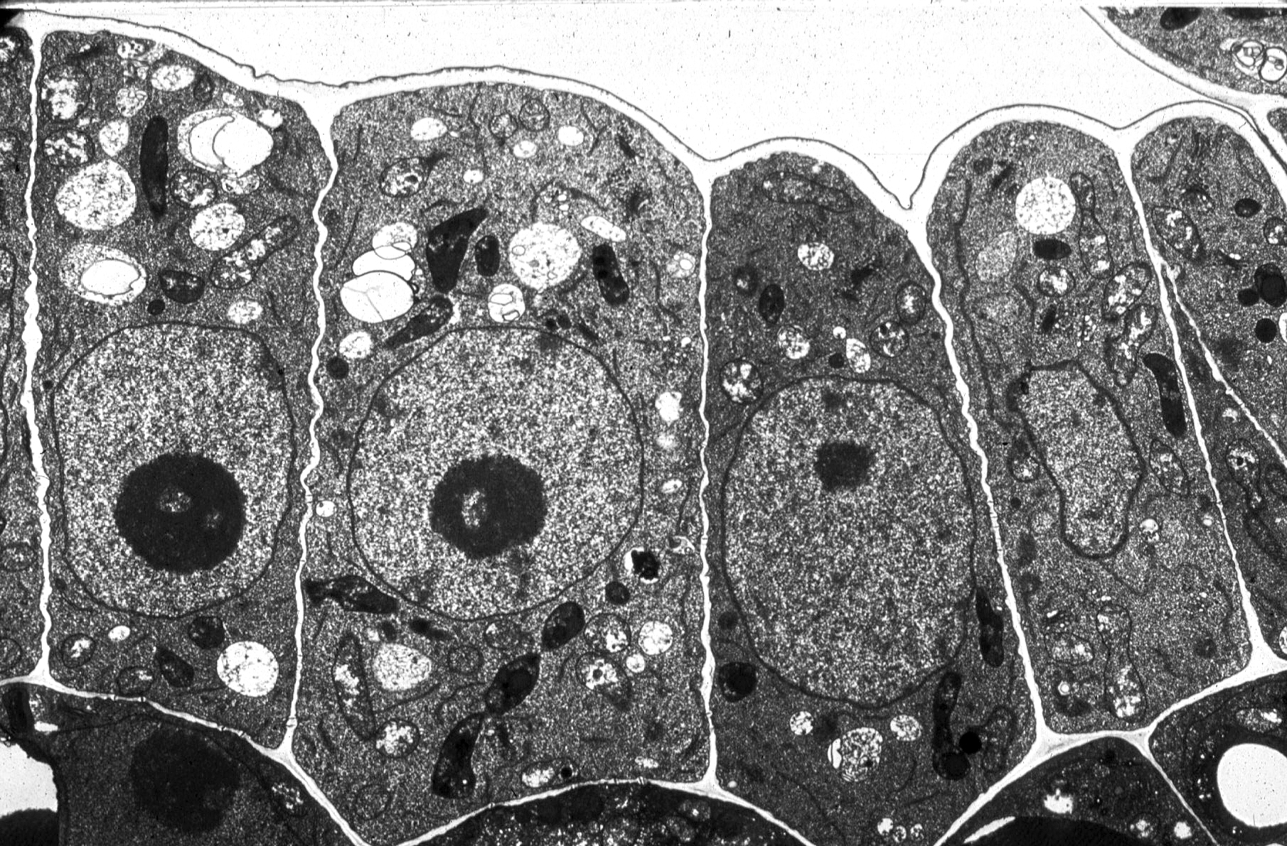
General structure of an animal cell as seen under a light ... from sites.google.com These represent as much as 8% of total cellular dry weight. Animal cells are of various sizes and have irregular shapes. We can say that cells are the basic structural and functional units of all living organisms. Animal cell under electron microscope. These cells come in all shapes and sizes and their structure adapts to their function. Electron microscopes use electron beams focused by electromagnets to magnify and resolve microscopic specimens. These are both specific types of cells, and from specific species. After observing the cyanobacterial cell under electron microscope, it appears multilayered present between the sheath and plasma membrane.
These are both specific types of cells, and from specific species.
Identify the different cell shape and relate it to the cell's function. Which of the following structures is found in animal cells. It separates cell organelles from cytosol. These cells come in all shapes and sizes and their structure adapts to their function. Animal cells are of various sizes and have irregular shapes. Pupil activity • cell structure • read through the information on each of the organelles as you colour them in • follow the guidance on colouring them in given at the bottom of the page • this works on the theory that whilst. These are both specific types of cells, and from specific species. These represent as much as 8% of total cellular dry weight. Animal cell under electron microscope. Under electron microscope, cell membrane appears trilaminar (made. Identify the basic structures of a cell 2. Under the microscope, an animal cell shows many different parts called organelles, that work together to keep the cell functional. A cell is a very tiny structure which exists in living bodies.
Share :
Post a Comment
for "Structure Of Animal Cell Under Electron Microscope : animal cell under a microscope - This is because of the way that the cell was sectioned (cut) before it was viewed on the transmission electron microscope."


Post a Comment for "Structure Of Animal Cell Under Electron Microscope : animal cell under a microscope - This is because of the way that the cell was sectioned (cut) before it was viewed on the transmission electron microscope."